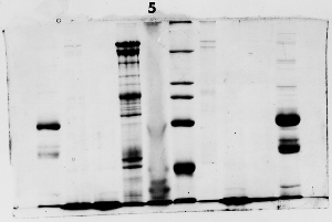
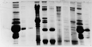
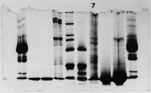
Overview: [Characterization of red cell membrane proteins by SDS-PAGE] [research paper]
Topics: [gel analysis] [molecular mass standard curve] [measuring relative mobility] ["Hall of Shame"]
Data: [gel images]

Streaked Protein Gels

Streaks – example 1

This one isn't so bad. The lane with the streaks (left of lane 5) is quite usable, in fact. The streaks came about because a portion of the more concentrated proteins in the membrane fraction precipated during the stacking process, then returned to solution after a fairly short time. Because of slight imperfections in the separating gel surface (it may have been allowed to dry a little before adding the stacking gel), the protein concentration was not uniform across the lane, and streaks rather than a uniform darker background are the result.

The electric field was quite uniform, and diffusion minimal, otherwise the streaks would not remain as narrow vertical lines.

Streaks – example 2

The fourth lane is streaked due to the effect described above. The electrical field was affected by the non-uniform concentration in the fourth lane (the dark one with streaks),, causing a narrowing of the bands.

Note that the dye front was lost in this gel - it was allowed to run too long.

Streaks – example 3

This one kind of stinks. Evidently, this group did not use an overlay after pouring the first gel layer. The result was a separating gel with the very rough top that you see here. As samples were stacked they were disproportionately concentrated in the 'valleys,' where the proteins precipitated. The precipitates re-dissolved gradually during the run, forming streaks instead of resolving into discrete bands.


Copyright and Intended Use
Visitors: to ensure that your message is not mistaken for SPAM, please include the acronym "Bios211" in the subject line of e-mail communications
Created by David R. Caprette (caprette@rice.edu), Rice University 9 Oct 96
Updated 26 May 05