Home
Studies
& Data Analysis
Methods
Microscope studies
Flagella experiment
Laboratory math
Blood fractionation
Gel electrophoresis
Protein gel analysis
Mitochondria
Concepts/ theory
Keeping a lab notebook
Writing research papers
Dimensions & units
Using figures (graphs)
Examples of graphs
Experimental error
Representing error
Applying statistics
Principles of microscopy
Solutions & dilutions
Protein assays
Spectrophotometry
Fractionation & centrifugation
Radioisotopes and detection
- overview; types; bright field microscopy
- dark field optics
- phase contrast
- oil immersion
- differential interference contrast
- measuring
- using a counting chamber
- wet (Vaseline) mount
Phase Contrast Microscopy
Most of the detail of living cells is undetectable in bright field microscopy because there is too little contrast between structures with similar transparency and there is insufficient natural pigmentation. However the various organelles show wide variation in refractive index, that is, the tendency of the materials to bend light, providing an opportunity to distinguish them.
A culture of Amoeba proteus or a fresh suspension of Nagleria gruberi make good practice specimens.
Principle
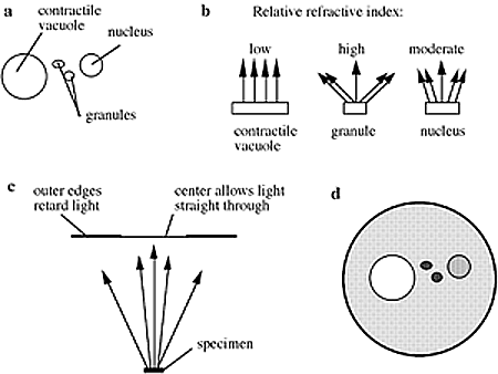
Highly refractive structures bend light to a much greater angle than do structures of low refractive index. The same properties that cause the light to bend also delay the passage of light by a quarter of a wavelength or so. In a light microscope in bright field mode, light from highly refractive structures bends farther away from the center of the lens than light from less refractive structures and arrives about a quarter of a wavelength out of phase.
Light from most objects passes through the center of the lens as well as to the periphery. Now if the light from an object to the edges of the objective lens is retarded a half wavelength and the light to the center is not retarded at all, then the light rays are out of phase by a half wavelength. They cancel each other when the objective lens brings the image into focus. A reduction in brightness of the object is observed. The degree of reduction in brightness depends on the refractive index of the object.
Applications for phase contrast microscopy
Phase contrast is preferable to bright field microscopy when high magnifications (400x, 1000x) are needed and the specimen is colorless or the details so fine that color does not show up well. Cilia and flagella, for example, are nearly invisible in bright field but show up in sharp contrast in phase contrast. Amoebae look like vague outlines in bright field, but show a great deal of detail in phase. Most living microscopic organisms are much more obvious in phase contrast.

Figure. (a) organelles are nearly invisible in bright field although they have different refractive indexes; (b) light is bent and retarded more by objects with a high refractive index; (c) in phase contrast a phase plate is placed in the light path. Barely refracted light passes through the center of the plate and is not retarded. Highly refracted light passes through the plate farther from center and is held back another one quarter wavelength.; (d) The microscope field shows a darker background (in this case the cell cytoplasm has a higher refractive index than the contractile vacuole), with the organelles in sharp contrast.
Using phase contrast
Phase contrast condensers and objective lenses add considerable cost to a microscope, and so phase contrast is often not used in teaching labs except perhaps in classes in the health professions and in some university undergraduate programs. This is unfortunate since the images obtainable in phase contrast mode can be very dramatic.
To use phase contrast the light path must be aligned. An element in the condenser is aligned with an element in a specialized phase contrast lens. This usually involves sliding a component into the light path or rotating a condenser turret. The elements are either lined up in a fixed position or are adjusted by the observer until the phase effect is optimized. Generally, more light is needed for phase contrast than for corresponding bright field viewing, since the technique is based on a diminishment of brightness of most objects.
Visitors: to ensure that your message is not mistaken for SPAM, please include the acronym "Bios211" in the subject line of e-mail communications
Created by David R. Caprette (caprette@rice.edu), Rice University 11 May 00
Updated 10 Aug 12
