Maneesh defended his PhD thesis in October, 2003. He now works on the artificial gravity project for Wyle Laboratories in Texas City, TX. His webpage while at Rice is provided below:
|
Initiation
of Platelet Thrombus Formation: Analysis Using Optical Tweezers
Optical tweezers are an extremely useful
tool in the fields of cellular
and
molecular biology. In the lab of Dr. Bahman Anvari, I use the
optical tweezers
to study intermolecular forces of various receptor-ligand interactions
associated
with the cardiovascular system. Thrombosis is one of the leading causes
of sickness and death and is clinically more important than all of the
hemorrhagic
disorders combined. Thrombi can form in any part of the
cardiovascular
system and appear due to some type of endothelial cell
perturbation.
While the endothelial cells are involved in the initial stage of
thrombus formation,
the main participants in thrombogenesis are the platelets.
Platelets
adhere, alter their shape, and secrete their granular contents in
thrombogenesis.
Numerous receptors on the surface of the platelet interact with various
ligands causing adhesion, platelet aggregation, and a change in
platelet shape.
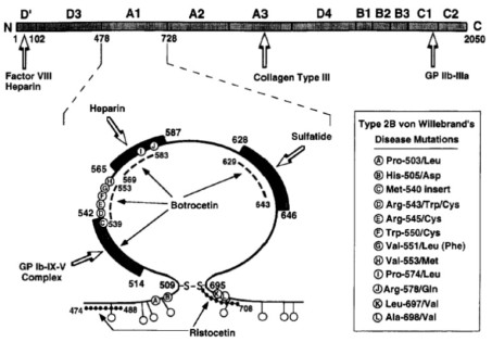
The glycoprotein (GP) Ib-IX-V complex is the protein on the platelet
surface
that initiates thrombus formation. GP Ib-IX-V mediates the
deposition
of platelets on the subendothelium in a damaged area and initiates
thrombosis
by binding under high shear conditions (as in regions of a damaged
vessel) to a
multimeric protein called von Willebrand factor (VWF) that circulates
in the plasma.
By quantitatively evaluating the VWF/GP Ib-IX-V bond strength using
optical
tweezers, we can gain new insight into the individual molecular
interactions
involved in thrombogenesis and eventually develop new drugs that target
specific
stages in the formation of thrombi.
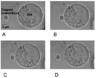
Using our optical tweezers, I examine the intermolecular force between GP Ib-IX-V and VWF. As a model system, we use transfected Chinese hamster ovary cells that express the platelet glycoprotein receptor. In addition, we use polystyrene beads coated with VWF. In our experiments, we trap the coated bead and let the bead and the cell adhere for a certain amount of time. We then use a piezo-electric stage to detach the cell from the bead. Using Stokes's Law, we can compute the binding force between VWF and GP Ib-IX-V. We use a titanium sapphire laser tuned to a wavelength of 830 nm to minimize cellular damage. Multimers of VWF that are of average size do not normally bind platelets so we use the exogenous modulators ristocetin and botrocetin to induce VWF/GP Ib-IX-V adhesion. Our results are very interesting because we notice that the detachment forces of the bead from the cell occur in quantal units. Other groups, which investigate different receptor-ligand interactions using optical tweezers, also frequently observe discrete force quantities associated with the rupture of bonds. I have also examined the bindi color(255,255,255);ng of unusually large forms of VWF to GP Ib-IX-V and the role that various amistyle="top: 0px; height: 1600px;"no acids of the platelet receptor complex play in adhesion. In future, I plan to investigate adhesion of different mutants of GP Ib-IX-V to VWF and also determine the significance of loading rate when detaching the bead from the cell. |
|
Research
Interests: Intermolecular
forces, optical tweezers, platelet adhesion
|

|
Maneesh
Arya
|
|
Bead
adhesion and subsequent detachment from CHO cell. (A) VWF-coated bead
is optically
trapped, and CHO abIX cell is moved toward towards bead. (B) Bead
adheres
to cell for 10 sec. (C) After increasing laser power, bead begins to
detach.
(D) Bead is fully detached.
|
|
Copyright
©2002
Photomedicine and Biomedical Photonics Lab Maintained by Brian Pikkula Last Update: July, 2002 |