

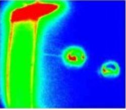
Electromotility and Membrane Electromechanics
Using a novel instrument that
combines optical trapping with voltage clamping and fluorescence
imaging, we are studying the
electromechanical properties of
the cochlear outer hair cells (OHCs) plasma membrane in
collaboration with Dr. William Brownell in the
Department of
Otorhynolaryngology at Baylor College of Medicine, Doctors Aleksander
Popel and
Alexander Spector in the Department of Biomedical
Engineering at Johns Hopkins University, and Dr. Robert Raphael in the
Department of Bioengineering. The OHCs are the only known biological
materials capable of
producing
electrically-evoked forces, known as electromotility, over a wide range
of frequencies (up to at approximately 50 kHz and possibly higher).
Characterizing the electromechanical properties of the OHCs plasma
membrane, and understanding the contribution of prestin to
electromotility not only has implications for understanding the normal
hearing process and strategies for treatment of specific types of
hearing
loss, but also direct relevance to design and development of membrane
(lipid) -based micro/nano-electromechanical devices that could
potentially be used for diagnostic and therapeutic purposes.
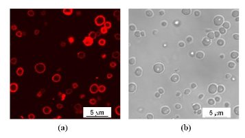
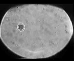
Chromophore-encapsulated Nano-Assembled Complexes for Optical Therapy
We are also developing new techniques to improve the efficacy of optical treatment for various types of tissue anomalies. The current therapeutic approach used in clinics relies on the selective absorption of laser irradiation to elicit spatially-confined thermal damage. Through collaborations with Dr. Michael Wong in the Department of Chemical and Biomolecular Engineering, we are investigating the utility of novel microcapsules containing chromophores to serve as exogenously administered optical targets. These capsules allow for the localized delivery of chromophores sensitive to near infrared wavelengths, which penetrate deeper within tissue and are absorbed less by interfering intrinsic chromophores such as melanin.