

Kathryn defended her PhD thesis in January, 2004. She now works as a medical writer for Medtronic Sofamor Danek. Her webpage while at Rice is provided below:
|
Quantification
of Staphylococcal Adhesion Using Optical Tweezers
Biofilm
formation, a common cause of medical device failure, initially results
from
bacterial adhesion to an adsorbed layer of extracellular matrix (ECM)
proteins
on the implant surface. Molecules called microbial surface
components
recognizing adhesive matrix molecules (MSCRAMMs) on the surface of
bacterial cells
mediate this adhesion. Because an early step in biofilm
development is
the binding of bacteria to an adsorbed protein layer, developing a
greater understanding
of the interactions between MSCRAMMs and their ligands can lead to
improved methods of preventing bacterial adhesion or removing adherent
bacteria.
The objective of my project is to use optical tweezers to characterize
various aspects of MSCRAMM-related staphylococcal adhesion to surfaces
coated with
extracellular matrix proteins.
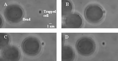
Optical tweezers, which are based on Maxwell's theory of radiation pressure, use a highly focused laser beam to exert force on microscopic objects. To study bacterial adhesion, we use optical tweezers to detach bacterial cells from polymer microspheres coated with ECM proteins. We can then determine the amount of force required for this detachment. The process of adhesion and detachment is illustrated in the micrographs to the right. |

|
Kathryn
Simpson
|
|
The
bacterium is trapped (A) and held against a coated microsphere for 10
seconds
(B). The trapping power is increased to initiate the
detachment process
(C), and finally the cell completely detaches from the microsphere (D).
|
|
Copyright
©2002
Photomedicine and Biomedical Photonics Lab Maintained by Brian Pikkula Last Update: July, 2002 |