
The Cerebral Hemispheres
The Telencephalon (the cerebral hemispheres) is the largest of the divisions of the human brain, and it is what subserves language — at least the aspects of language which are of interest to linguistics and most other people. In fact, the same can be said of the just the cerebral cortex, only one of the four parts of the telencephalon according to the traditional division given previously.
Of the subcortical and interior portions of the telencephalon, the basal ganglia, which partially surround the diencephalon, participate in motor functions, including articulation of speech, and the hippocampus and the amygdaloid nucleus, which lie deep within the lower part of the cortex, are very important in emotional expression.
The hippocampus is actually a paleocortical structure (thus part of the cortex), deeply embedded within the lower temporal lobe of either hemisphere. If is important for remembering events of one's personal history and other kinds of what are known as declarative memory, and so it looks large in linguistic activity, as the linguistic system clearly has access to this kind of information- it is an important part of semology. More than that, it appears that the hippocampus, since it plays an important role in declarative memory (along with higher cortical levels), is of great importance in language learning of the kind commonly done in high schools and colleges, which is quite different from that done by children, who automatically use lower levels of their cerebral cortices, acquiring their native language skills as procedural knowledge rather than declarative knowledge. That difference is directly correlated with the great skill and fluency of people using their native languages as opposed to the clumsiness with which they attempt to navigate their way in a foreign country with the aid of a school-taught second language.
The Cerebral Cortex
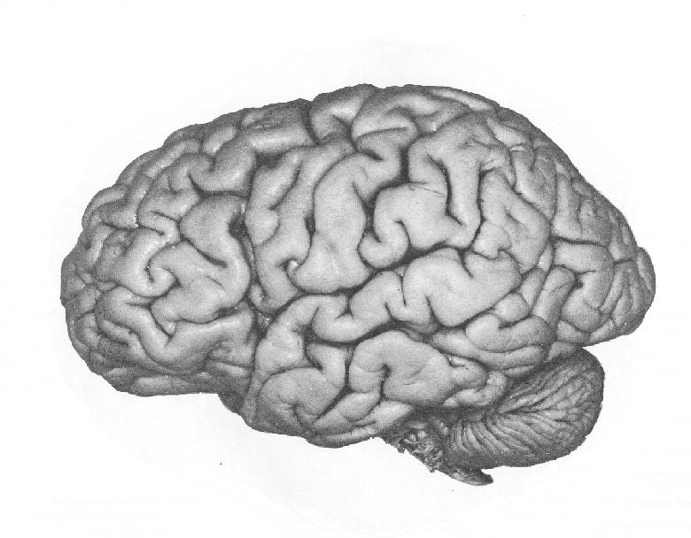
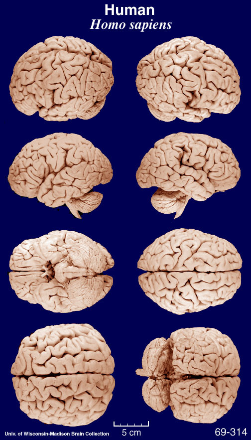
The cortex is what you see in popular pictures of the brain, and it is all you see in most of them unless they happen also to show the cerebellum, as a few do. It is characterized visually by its numerous grooves; and it was once supposed by many that the function of these grooves was somehow to store information, in much the same way that the grooves of a phonograph record store information. It is a good example of how people use the familiar as a metaphor for attempting to grasp the unfamiliar — usually with significant lack of success, since the currently popular technology in any era is rarely if ever much like that of the brain. This metaphor was popular back in the days of phonograph records. Luckily, we are now beyond that era; unluckily, we are now in the computer era, which provides a metaphor only slightly better and almost as misleading, but one to which many cognitive scientists, people who you might think would know better, have given credence.
What is the real reason, then, for
all those grooves? To understand the answer to this question, it is helpful
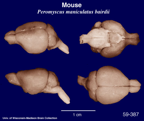
to look at the brains of some other mammals. The rat's cortex is very small
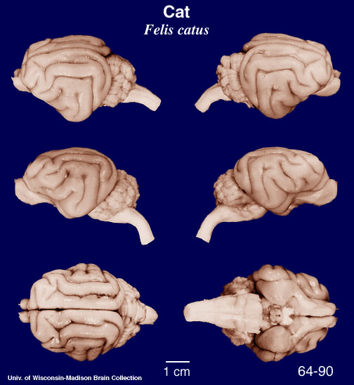
in its horizontal spread and has practically no grooves; the cat's cortex
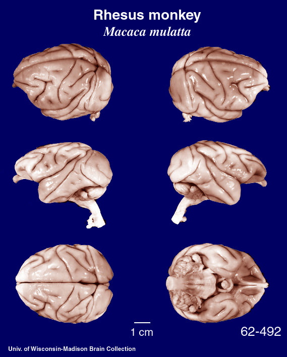
is larger with a little folding; and the monkey's larger still with yet
more grooves, including a Sylvian fissure and a central sulcus (see below
for these terms) and a few more. We find that the cortices of different
mammals differ not only in size but also in the extent of folding —
the larger the cortex, the more grooves —
but there is not much difference in
the thickness of the cortex from one mammal to another, despite the enormous
difference in overall brain size. To get an idea of the latter, the cortex
of the cat has been estimated to be about seventy times as large as that
of the mouse, and that of the human about 3400 times as large.
(Click on any of the pictures below for an enlarged view.)
 -
- -
- -
-
But the thickness among these different animals' cortices varies only from about one or two millimeters in mouse cortex to around three to six millimeters in human cortex. Within that thickness can be identified six layers of structure, and in this property also we find great uniformity among different mammals.
What appears to have happened during the long process of evolution of the human brain is that the cortex has grown, but topologically the growth is almost entirely in just two of the three spatial dimensions and not in thickness. If you could unfold it, eliminating all those hills and valleys, the resulting form would be very thin in one dimension (three to five mm) with great extent in the other two, amounting to a surface area of about 1400 square centimeters or one-and-a-half square feet. In the course of evolution, it has spread out enormously while acquiring very little additional thickness. And the only way to get such a large surface area inside a skull small enough to allow for childbirth is to crumple it up. That is the reason for all those grooves. Another is that the connections from one part of the cortex to another, which pass through the white matter, for example between frontal and posterior areas and between the left temporal and right temporal lobes, can be much shorter than they would have to be without all that folding.
Hemispheres and Lobes
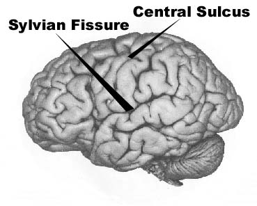
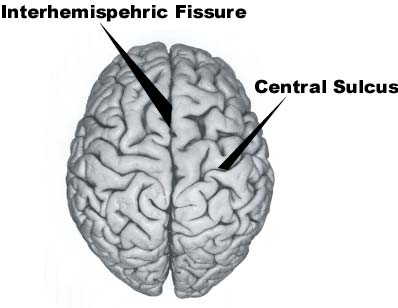
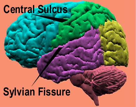
What has been called a groove above is actually called a Sulcus (plural 'sulci') or a Fissure, depending on its size; the two really large and deep ones are usually called fissures (but one of these, the Sylvian fissure, is sometimes called the lateral sulcus). These two fissures plus the Central Sulcus(a.k.a 'Rolandic fissure') divide the cortex into its main divisions. The most prominent is the Interhemispheric Fisssure (a.k.a. 'longitudinal fissure'), which divides the cerebral cortex into two hemispheres, right and left. The two hemispheres are similar in structure if not looked at too closely, different in details; but both have the same major partitions into lobes and their subdivisions.
 ---
---
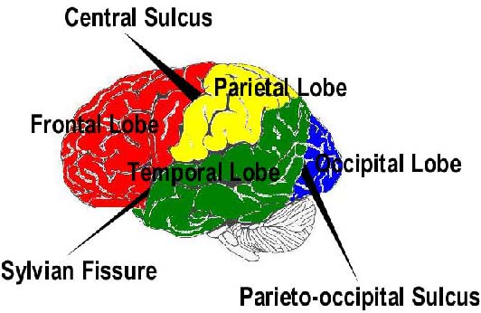
The next most prominent feature is the Sylvian Fissure(a.k.a lateral sulcus). Below the Sylvian fissure is the Temoral Lobe. Next most prominent is the Central Sulcus; the traditional name, still used sometimes, is 'Rolandic Fissure'. Anterior to this sulcus is the Frontal Lobe. Posterior to it and above the Sylvian Fissure, we have the Parietal Lobe and the Occipital Lobe. And what separates these two from each other? One can say it is the parieto-occipital sulcus, and that would be correct, but you won't see it in lateral views of the cerebral hemisperes because it is only apparent from inside the interhemispheric fissure. Originally, the parietal lobe was distinguished and so named because it is the part of the brain just beneath the parietal bone; and the occipital lobe is just beneath the occipital bone. These bones don't fuse until some time after childbirth, allowing the skull to be a little flexible for that process, of great benefit in the trade-off between having room there for all those billions of neurons and having too much brain volume for passage through the birth canal.
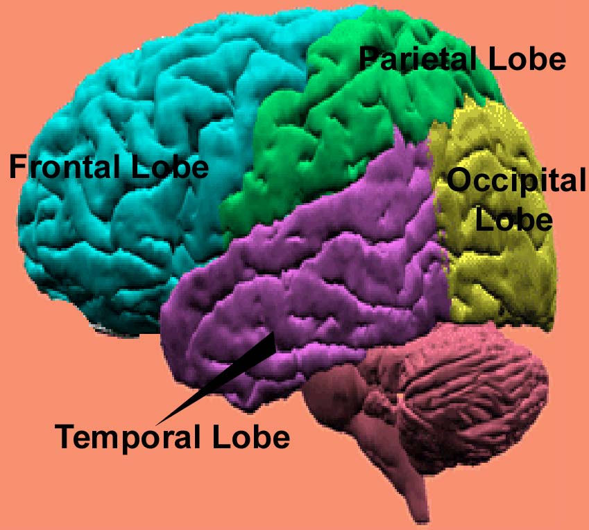
These then are the four lobes: frontal, temporal, parietal, occipital. Their names are pretty straightforward: the frontal lobe is in front; the temporal lobe underneath the temple; the parietal and occipital lobes are named for their overlying bones, and these bones get their names from Latin: 'parietal' comes from the word for 'wall'; 'occipital' is from ob- "in back of" and caput "head", with assimilation of the b to the following c: "at the back of the head".
Subdividing the Lobes
All of the lobes are further subdivided by smaller sulci. For our purposes we need to consider only a few of them. Between the sulci are Gyri (singular 'gyrus'); it is mainly the gyri that you see in pictures of the surface of the cortex. If the sulci are like valleys and canyons, the gyri are like (long) hills and ridges.
See the appropriate lobe page for further subdivisions:
There is yet another
complicating factor, a very important one: Different people have
different brains (hardly a surprise). Just as some people have long legs,
others short ones; some have big noses; some have high cheek bones; some
have big ear lobes, others tiny ones; so also we find great differences
in the details of location of the various sulci and gyri. In some brains,
the posterior portion of the Sylvian fissure makes a sharper upward turn
than in others; in some the central sulcus is not as vertical as in others;
and so forth.
Go to Menu (for non-frame version.)
© 2000 Rice University. This document, or any portion hereof, may be used for non-commercial informational purposes only. Any copy of this document, or portion hereof, must include the copyright notice (http://www.rice.edu/about/cr-notice.html) in its entirety.